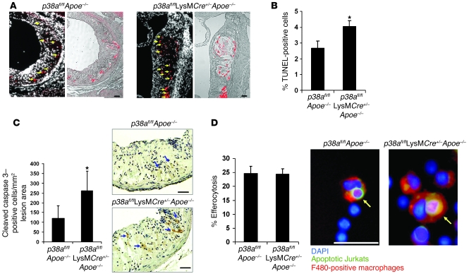
Figure 3. Increased apoptosis in plaques deficient in macrophage p38α MAPK.
(A–C) Sections from the proximal aortas of p38afl/flApoe–/– and p38afl/flLysMCre+/–Apoe–/– mice were labeled by TUNEL to detect apoptotic cells and counterstained with DAPI to detect nuclei, or stained for activated caspase 3. (A) Shown are TUNEL-positive cells (red) in the intimal area that colocalized with DAPI-stained nuclei (arrows; left) or necrotic area (right). (B) Mean percent TUNEL- and DAPI-positive cells relative to DAPI-positive cells in the lesion (n = 9 per genotype). (C) Number of cleaved caspase 3–positive cells per unit of lesion area (n = 7 per genotype). Shown are representative images of activated caspase 3 staining in the intimal area of the lesions. Arrows indicate caspase 3–positive cells. (D) In vivo efferocytosis assay. Shown are representative images of an efferocytic event that was counted as positive. Data are expressed as the percent of F4/80-positive macrophages (red) that had ingested an apoptotic T cell (green). See Methods for details. Shown are representative images of macrophages that were counted positive for efferocytosis (arrows). Approximately 200 F4/80-positive macrophages were counted per group. *P < 0.05 versus p38afl/flApoe–/–, Mann-Whitney U test. Scale bars: 50 μm.

