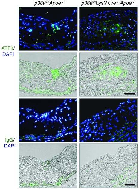
Figure 5. Induction of ER stress in vivo.
Aortic sections from p38afl/flApoe–/– and p38afl/flLysMCre+/–Apoe–/– mice were stained by immunofluorescence with an antibody against ATF3 or an IgG control antibody. ATF3-positive cells (green) in the intimal area (green staining overlaid on bright field) colocalized with and around the DAPI-stained nuclei (green staining overlaid on DAPI). Scale bars: 50 μm.

