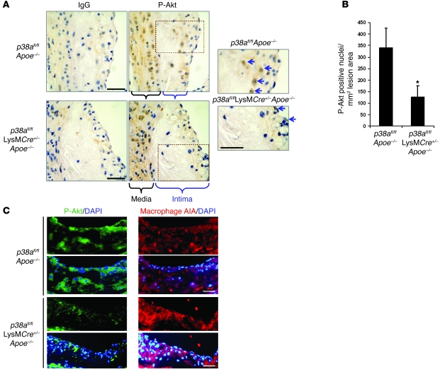
Figure 8. Akt phosphorylation is suppressed by macrophage p38α deficiency in vivo.
(A) Sections from the proximal aorta were stained for immunohistochemistry with an antibody against activated phosphorylated Ser473-Akt or an IgG control antibody. Medial and intimal areas are indicated. Areas within dotted outlines were enlarged to show the phosphorylated Akt–immunopositive nuclei in the intimal area (arrows). (B) Immunopositive nuclei were quantified in the intimal areas of the lesions from duplicate stained sections and averaged. Data are expressed as the total number of immunopositive nuclei per total lesion area (n = 4 per genotype). *P < 0.05 versus p38afl/flApoe–/–, Mann-Whitney U test). (C) Mouse lesions were also stained for phosphorylated Akt by immunofluorescence or for macrophages in adjacent sections. Shown are representative images of phosphorylated Akt (green) or macrophage staining (red) overlaid with DAPI-stained nuclei. Scale bars: 50 μm.

