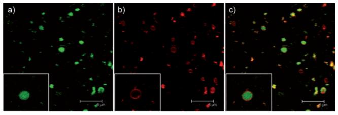
Fig. 1.

Confocal microscope images of green-fluorescent PA encapsulated by red-fluorescent asolectin liposomes. a) Green channel, b) red channel and c) overlaid images of the green and red channels. Insets show details of an individual liposome. Scale bar = 6 μm.
