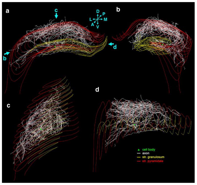
Fig. 3.

NeuroLucida reconstruction of the CA3 pyramidal cell. a Coronal view of the entire axon arbor. Green triangle shows the location of the cell body, the axons are white, the dentate granule cell layer is marked with yellow line, CA1-3 pyramidal cell layer with red line. D dorsal, V ventral, L lateral, M medial, A anterior, P posterior. b This rotated view shows that CA3 pyramidal cell axon collaterals follow the curve of the cornu Ammonis. c View of the axon arbor from dorsal. d View from medial
