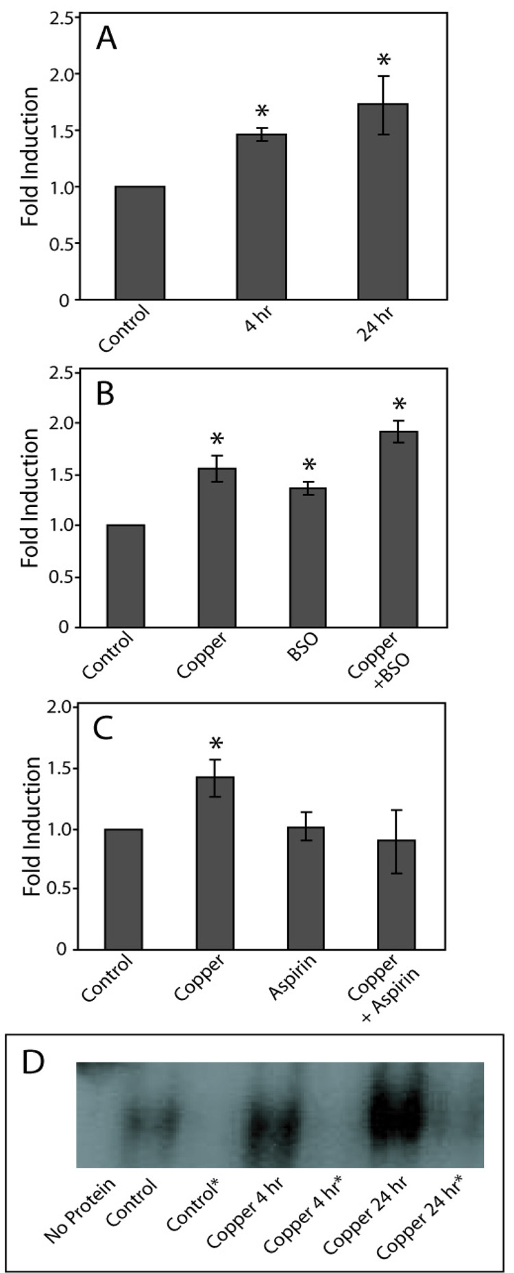
Figure 2. Effect of copper on binding to AREs.
COS-7 cells were exposed to 400 µM copper 4 or 24 h, and EMSA performed as described (panel A). In BSO experiments cells were pretreated with BSO prior to the addition of copper (panel B). In aspirin experiments, cells were pre-treated with aspirin prior to copper exposure (panel C). Data are expressed as mean fold induction +/− S.E.M. in comparison to untreated controls. * indicates significantly different from untreated controls by ANOVA, p < 0.05, n = 3 observations. Panel D, Representative EMSA of the data presented in panel A, *indicates reactions that contained excess non-radiolabeled ARE-containing oligonucleotide.

