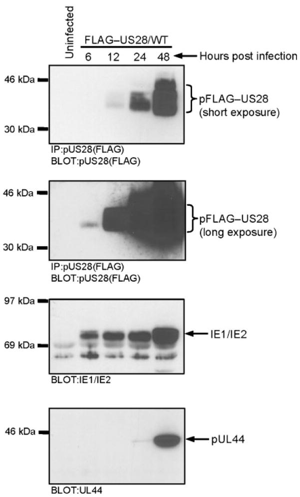
Fig. 3.
Kinetics of pUS28 expression in HCMV FLAG–US28/WT-infected cells. The pFLAG–US28 protein was immunoprecipitated from HCMV FLAG–US28/WT-infected HFFs (using anti-FLAG M2 agarose beads) and analysed by Western blotting using an α-FLAG polyclonal antibody. Overexposure of the blot enables pFLAG–US28 expression to be detected at 6 h post-infection (upper panels). Whole-cell lysates from the same time points were separated by SDS-PAGE and analysed by Western blotting using an α-IE1/IE2 antibody (lower middle panel) or an α-UL44 antibody (lower panel). Results shown are representative of three independent experiments.

