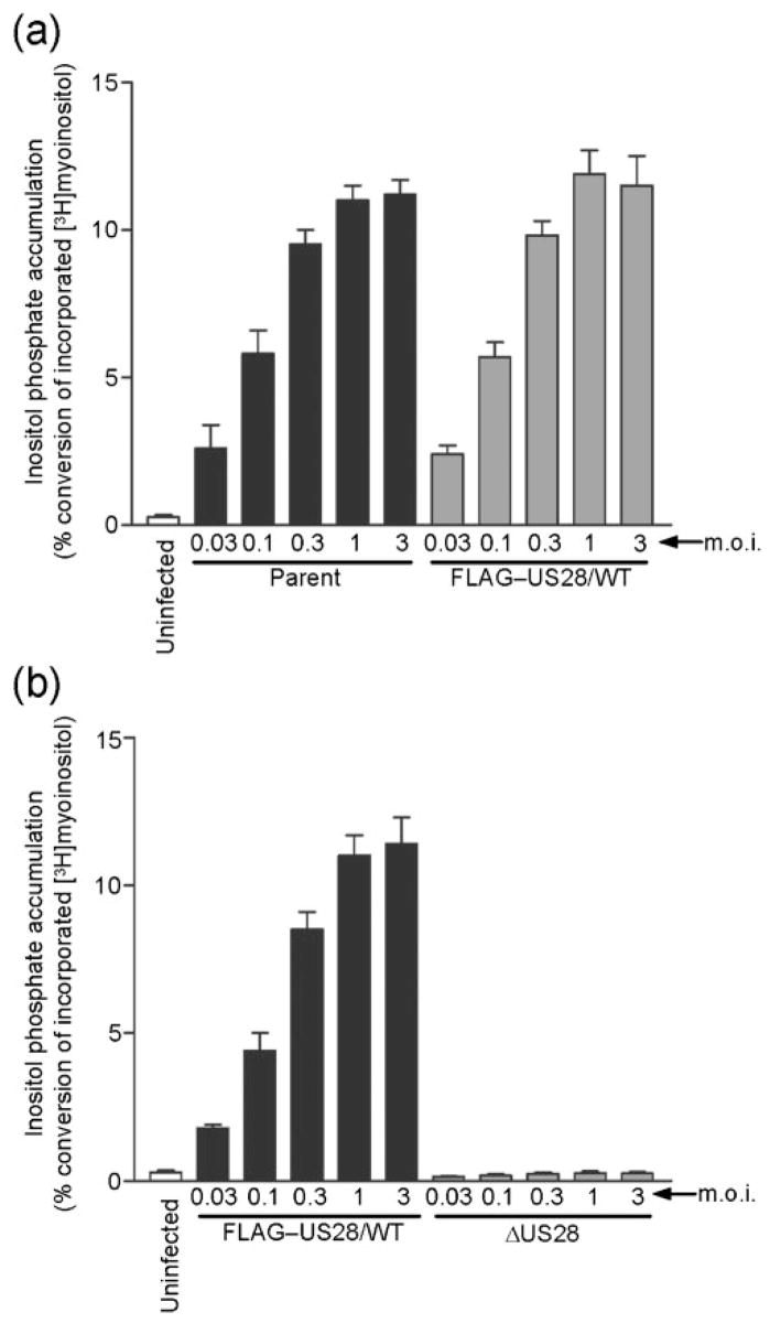
Fig. 5.

Characterization of PLC-βsignalling in cells infected with HCMV FLAG–US28/WT or HCMV ΔUS28. HFFs were infected with increasing m.o.i. (0.03 to 3.0 p.f.u. per cell) using HCMV parent or HCMV FLAG–US28/WT (a), and HCMV FLAG–US28/WT or HCMV ΔUS28 (b). Medium containing 1 μCi ml−1 (37 kBq ml−1) [3H]myoinositol was added at 24 h post-infection and accumulated InsPs were determined at 48 h post-infection using anion-exchange chromatography. The data represent four independent experiments performed in duplicate and are presented as a ratio of accumulated inositol phosphates to total 3H incorporation.
