
Fig. 3.
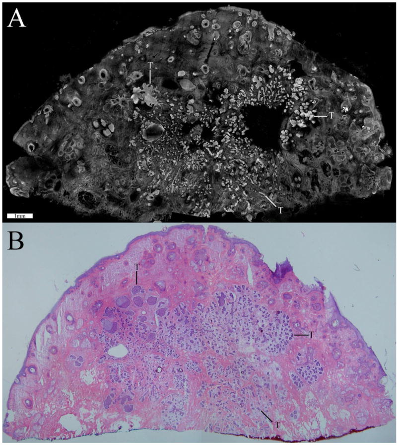
Confocal mosaic (a) of a micronodular BCC that compares well to the corresponding histology (b). Small and tiny nodules or nests of tumor are seen (T) appearing bright in the mosaic and purple-stained in the histology. (Color online only.)
Official websites use .gov
A
.gov website belongs to an official
government organization in the United States.
Secure .gov websites use HTTPS
A lock (
) or https:// means you've safely
connected to the .gov website. Share sensitive
information only on official, secure websites.
Confocal mosaic (a) of a micronodular BCC that compares well to the corresponding histology (b). Small and tiny nodules or nests of tumor are seen (T) appearing bright in the mosaic and purple-stained in the histology. (Color online only.)