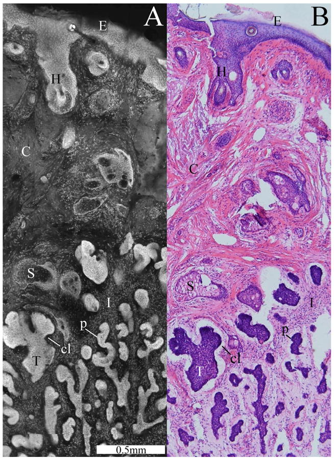
Fig. 4.

Confocal submosaic (a) showing a magnified 6× view of the mosaic in Fig. 3 and comparable histology (b). A micronodular BCC tumor (T) is seen with features such as nuclear pleomorphism, increased nuclear density, palisading (p), and clefting (cl). Normal features are seen such as the epidermis (E), a hair follicle (H), sebaceous glands (S), dermal collagen (C), and inflammatory cells (I).
