
Fig. 5.
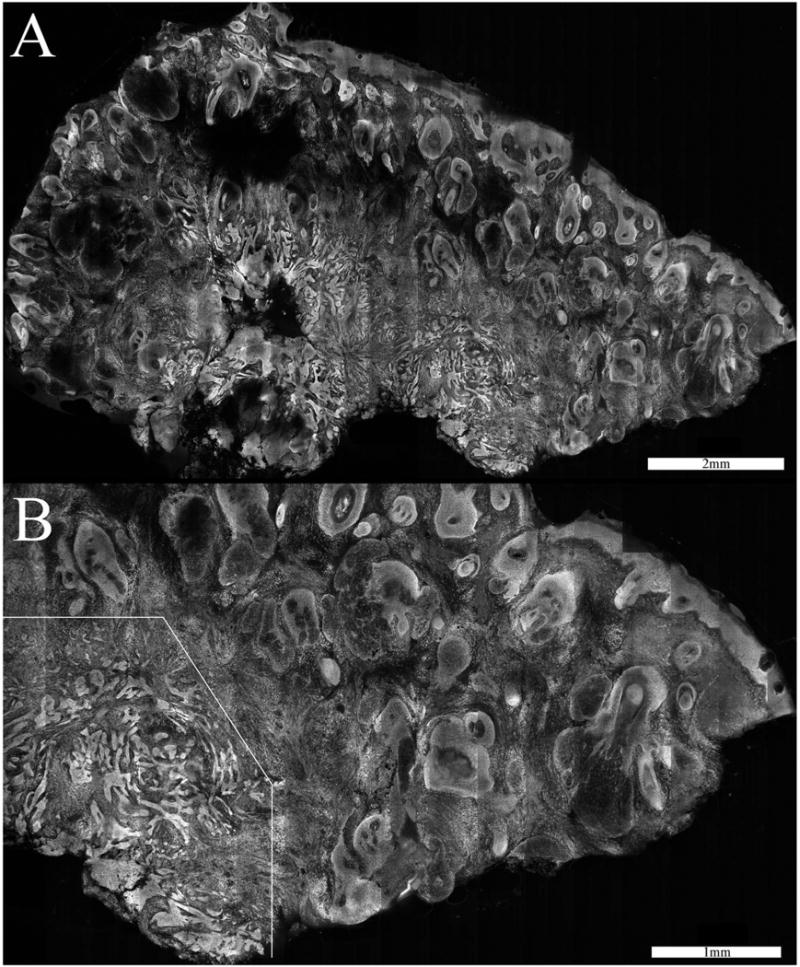
Confocal mosaic (a) of an infiltrative BCC. Thin strands of tumor lie in the deeper dermis. The tumor is not clearly visualized in the 2× view but is more obvious in the slightly magnified 4× view (b). The overall disruption of the tissue is evident in the lower-left portion.