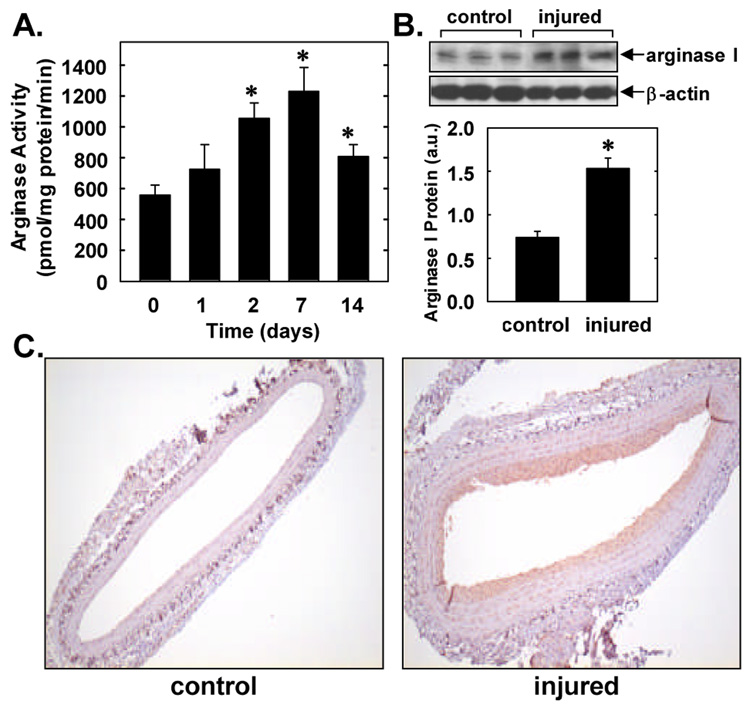
Figure 1. Effects of arterial injury on arginase activity and expression.
A. Arginase activity in rat control or balloon injured carotid arteries. Results are mean ± SEM of 5 experiments. *Statistically significant effect of arterial injury. B. Western blot of arginase I protein in control carotid arteries or in vessels 7 days post-injury. Arginase I protein levels were quantified by scanning densitometry, normalized with respect to β-actin, and expressed in arbitrary units (a.u.). Results are mean ± SEM of 3. *Statistically significant effect of arterial injury. C. Immunohistochemistry demonstrating arginase I expression (shown in brown) in control carotid arteries or in vessels 7 days post-injury (magnification 100x). Data are representative of 5 separate experiments.

