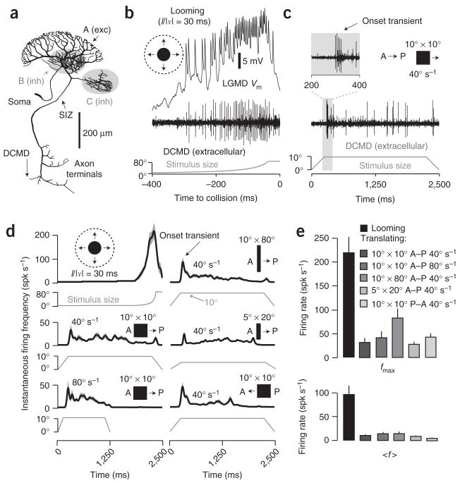
Figure 1.
LGMD morphology and response to looming versus translating stimuli. (a) LGMD reconstruction17 showing excitatory (A; exc) and inhibitory (B, C; inh) dendrites. (b) Response to looming (l/|v| = 30 ms). Top, LGMD membrane potential (Vm). Middle, corresponding nerve cord recording showing DCMD spikes. Bottom, the approaching disk’s retinal angle as a function of time to collision (t = 0). (c) Response to a 10° × 10° square, translating at 40° s−1 in the A–P direction. The onset transient is magnified in the inset. Stimulus size is the retinal angle subtended in the azimuth direction. The initial size increase is due to the object’s moving onto the screen. (d) Responses to assorted stimuli. Dark lines, gaussian-convolved instantaneous firing frequency (spikes s−1); gray envelopes, s.e.m. (N = 25 trials, 5 per locust). Left column: a looming disk (l/|v| = 30 ms; top) and 10° × 10° squares translating at 40° s−1 (middle) or 80° s−1 (bottom) in the A–P direction. Right column: 10° × 80° (top) and 5° × 20° (middle) rectangles translating at 40° s−1 in the A–P direction and a 10° × 10° square translating at 40° s−1 in the P–A direction (bottom). (e) Peak (fmax) and mean (〈f〉) firing rate in response to visual stimuli. 〈f〉 for looming was for the last 500 ms of approach; 〈f〉 for translation was for the steady state period (Methods). Error bars, s.e.m. (N = 25 trials, as in d); spk, spikes.

