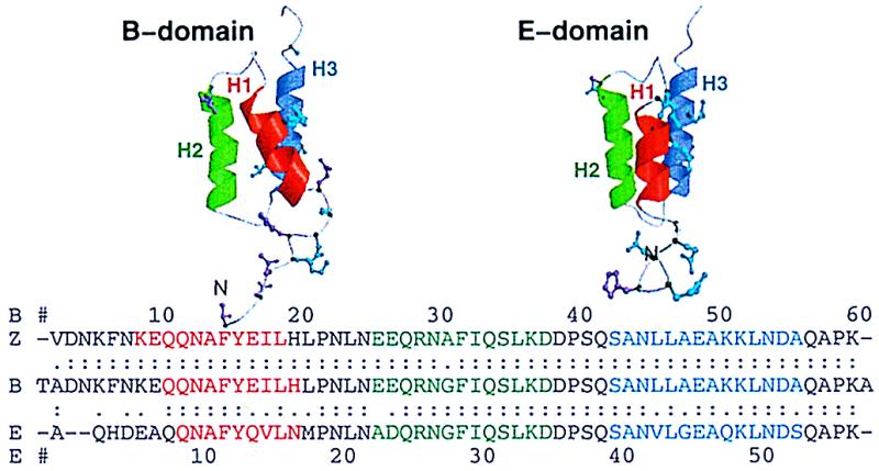
Figure 1.

Ribbon diagrams of the B and E domains and sequence alignment of the B, E, and Z domains of protein A. The helices are colored red, green, and blue for H1, H2, and H3, respectively, in both the structures and sequences. The helices in the E domain are as reported by Starovasnik et al. (ref. 6, Protein Data Bank ID 1edk): H1 (residues 8–15), H2 (22–34), and H3 (39–52). The helices in the B-domain NMR structure (ref. 8, 1bdd) are as follows: H1 (10–19), H2 (27–37), and H3 (42–55). The helices in the Z domain are as given by Tashiro et al. (ref. 9, 1spz): H1 (7–17), H2 (24–36), and H3 (41–54). Alignment was with the fasta algorithm (10). Identity is indicated with “:”. Conservative mutations are displayed in cyan and indicated with “.”. Nonconservative mutations are displayed in magenta and left blank in the alignment.
