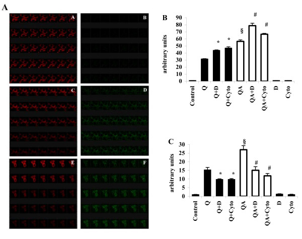
Figure 5.
Lipid peroxidation and ROS production in quartz-stimulated cells in the presence of DXS and Cytocalasin B. (A) Confocal microscopy analysis of lipid peroxidation of RAW 264.7 cells (single stacks acquired with oil objective 63.0 ×, digital zoom 8 ×) using the specific fluorescent probe BODIPY 581/591 in time-lapse experiments, with acquisitions of 1 slide/min. A, C, E show the decrease of red fluorescence during 35 min, while B, D, F show the contemporary increase of green fluorescence in the same cells. A, B control, untreated cells; C, D cells challenged with 100 μg/ml quartz particles; E, F cells challenged with quartz particles in the presence of 100 μg/ml DXS. (B) Fluorimetric quantitation of lipid peroxidation in RAW 264.7 cells challenged with 100 μg/ml Q or QA (white bars) particles in the presence (Q+D, QA+D and Q+Cyto, QA+Cyto) or absence of 100 μg/ml DXS or of 2 μg/ml Cytocalasin B for 1 h. Values are the mean ± SD from 4 experiments. The symbol § indicates a statistically significant difference between Q and QA challenged cells (T test, p < 0.0005); the asterisk indicates a statistically significant difference between Q+D or Q+Cyto and Q-challenged cells (T test, p < 0.005), while the symbol # indicates a statistically significant difference between QA+D or QA+Cyto and QA-challenged cells (T test, p < 0.005). (C) Fluorimetric measurement of ROS production in RAW 264.7 macrophages in the same conditions of (B) after 1 h incubation. Values are the mean ± SD from 4 experiments. The symbol § indicates a statistically significant difference between Q and QA challenged cells (T test, p < 0.0005); the asterisk indicates a statistically significant difference between Q+D or Q+Cyto and Q challenged cells (T test, p < 0.0025), while the symbol # indicates a statistically significant difference between QA+D or QA+Cyto and QA-challenged cells (T test, p < 0.0005).

