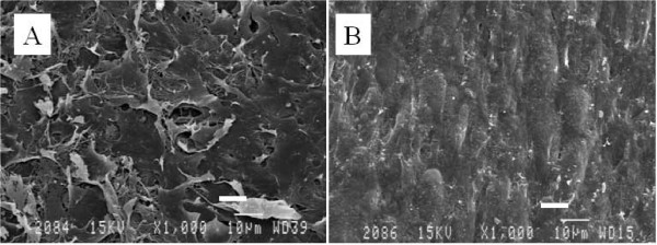
Figure 2.
Scanning electron microscopy. Scanning electron microscopy revealed that the top (A) and the basal aspects (B) of the chondrocyte sheet demonstrated completely different textures. The chondron like texture was only observed on the basal aspect, which had adhesive properties. Scale bar = 10 μm.

