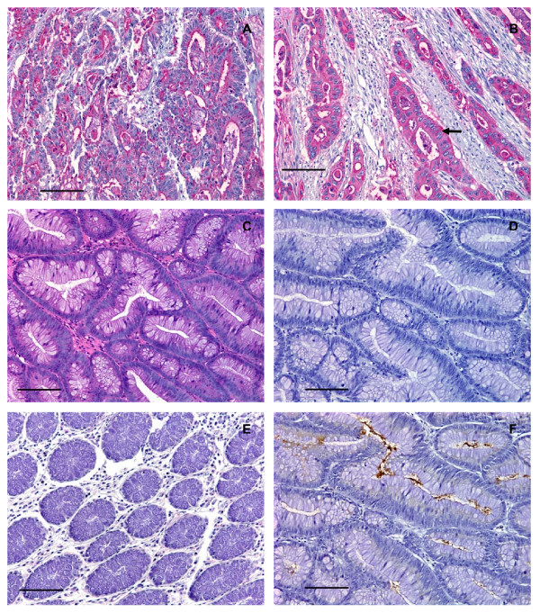
Figure 3.
Human colorectal adenocarcinomas, but not colorectal adenomas, expressed high levels of C2-O-sLex. (A) Photomicrograph of a tissue section of a moderately differentiated colorectal adenocarcinoma stained with CHO-131 mAb (15 μg/ml). Red color indicates positive cytoplasmic and luminal reactivity with CHO-131 mAb. (B) A poorly differentiated colorectal adenocarcinoma stained with CHO-131 mAb. Notice the red color indicating positive cytoplasmic reactivity of neoplastic cells (arrow). (C) A colorectal adenoma stained with hematoxylin and eosin. (D) A serial section of the same tissue as in (C), stained with CHO-131 mAb. Notice the absence of red color. (E) Mucosa of normal colorectal epithelium stained with CHO-131 mAb. (F) A serial section of the same tissue as in (C) and (D), stained with CEA mAb (1.6 μg/ml). The brown color indicates positive reactivity with CEA within the lumens of glands and in the cellular cytoplasm. All tissue sections were 4 μm thickness; scale bars = 100 μm, 200× magnification. Mayer's hematoxylin was used as a counterstain for tissues stained with CHO-131 and CEA mAbs. Representative sections from multiple stained tissues are shown.

