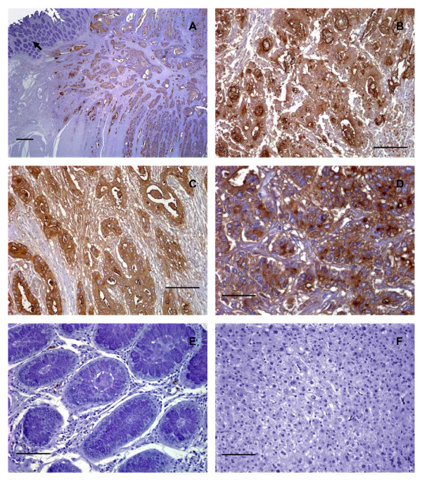
Figure 5.
Human primary colorectal adenocarcinomas and metastatic liver tumors expressed sLex. (A) Photomicrograph of a serial tissue section of the same well differentiated colorectal adenocarcinoma as shown in Figure 2A stained with CSLEX1 mAb (15 μg/ml). Brown color indicates positive reactivity with the CSLEX1 mAb. The arrow indicates adjacent normal colorectal mucosa that lacks reactivity with CSLEX1 mAb, scale bar = 500 μm. (B) A moderately differentiated colorectal adenocarcinoma stained with CSLEX1 mAb. Brown color indicates positive reactivity with CSLEX1 mAb, scale bar = 100 μm. (C) A serial tissue section of the same poorly differentiated colorectal adenocarcinoma as in Figure 3B stained with CSLEX1 mAb (15 μg/ml). Brown color indicates positive reactivity with CSLEX1 mAb, scale bar = 100 μm. (D) A moderately differentiated metastatic colorectal adenocarcinoma in liver stained with CSLEX1 mAb. The cytoplasm of neoplastic cells reacted positively with CSLEX1 mAb as indicated by the brown color, scale bar = 50 μm. (E) Mucosa of normal colorectal epithelium stained with CSLEX1 mAb. Note the absence of brown color, indicating a lack of reactivity with CSLEX1 mAb, scale bar = 100 μm. (F) Normal liver stained with CSLEX1 mAb. Note the absence of brown color, indicating a lack of reactivity with the CSLEX1 mAb, scale bar = 100 μm. All tissue sections were 4 μm thickness. Figure (A) 20× magnification, (B) and (C) 200× magnification, (D) 400× magnification, (E) and (F) 200× magnification. Mayer's hematoxylin was used as a counterstain. Representative sections from multiple stained tissues are shown.

