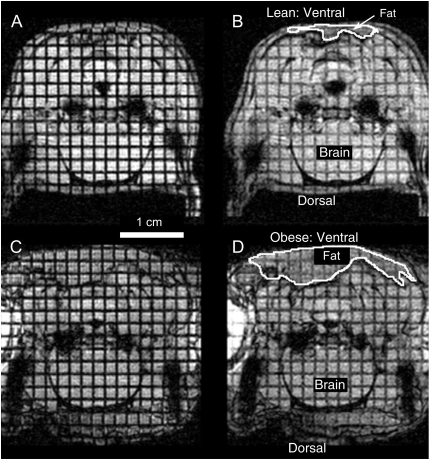
Figure 2.
Representative MR axial images of the hypopharynx (level 8) in a spontaneously breathing lean Zucker rat (A and B) and obese Zucker rat (C and D). Images in A and C were acquired during midexpiration; images in B and D were acquired just before the nadir of inspiration. The airway is the black area at the midline of each image. The black spaces lateral to the airways in each image are part of the tympanic bulla. Noninvasive grid lines were applied before the expiratory images (A and C) were acquired. Displacement of the grid lines in the subsequent inspiratory images (B and D), particularly in the lean Zucker rat, reveal the tissue strain in the pharyngeal walls. The grid lines provide fiducial markers that were used in the subsequent analysis of tissue strain. Annotations include the following: the 1-cm scale below A, and ventral and dorsal directions indicated for the lean rat in B and similarly for the obese rat in D. Approximate segmentation of typical fat regions in images of the lean rat (B) and obese rat (D) are outlined. The brain region is also noted in B and D.

