
Figure 5. Role of FOXO1 in PsA-induced apoptosis.
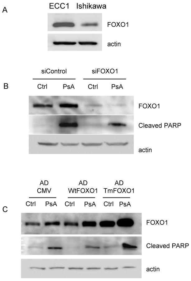
A) Ishikawa and ECC1 cells lysates were run on Western blot for FOXO1 and actin. B) ECC1 cells were transiently transfected with siRNA to FOXO1 (siFOXO1) or to a control luciferase gene (siControl). Cells were then treated with 1uM PsA for 24h. Western blots for FOXO1, cleaved PARP and actin were done. C) Ishikawa cells were infected with adenoviruses containing constructs for empty CMV (AD-CMV), wild type FOXO1 (WtFOXO1) or the triple mutant FOXO1 (TmFOXO1) cDNAs. Cells were then treated with 1uM PsA for 24h. Western blots for FOXO1, cleaved PARP, and actin were done. Data are representative of at least three independent experiments.