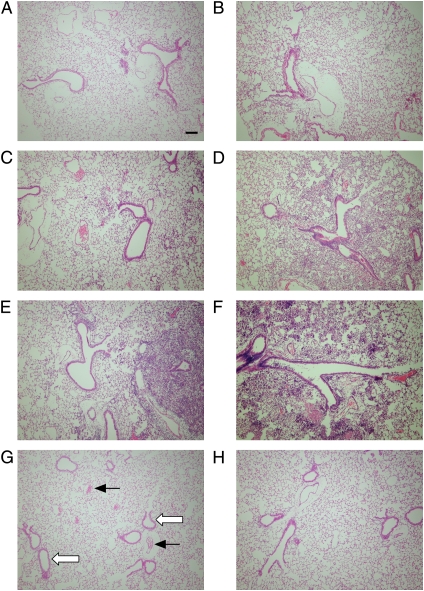
Figure 5.
Histology sections representing areas of inflammation in distal airways with adjacent alveoli after a single inoculation with P. aeruginosa. Wild-type (A, C, and E) and CF (B, D, and F) mice were inoculated with PA M57–15 by insufflation (∼ 5 × 10⁷ cfu/mouse), and killed 3 h (A and B), 1 d (C and D), or 2 d later (E and F; n = 6/group). Control wild-type and CF mice (G and H, respectively) were untreated. After BAL, the lungs were prepared for histologic examination, and stained with hematoxylin and eosin. Examples of blood vessels (solid arrows) and airway epithelia (open arrows) are indicated in an untreated wild-type mouse (G). Original magnification, 10×; bar = 100 μm.

