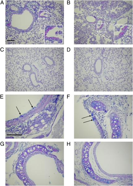
Figure 9.
Lung histology sections after repeated administrations of P. aeruginosa. Wild-type (A and E) and CF (B and F) mice were inoculated intranasally with P. aeruginosa on Day 0 (2.6 × 10⁷ cfu) and Day 2 (6.9 × 10⁸ cfu), and killed on Day 4. Lung sections from untreated wild-type (C and G) and CF (D and H) mice in similar areas of the lung are shown for comparison. BAL was performed and the lungs were prepared for sectioning and stained with alcian blue and periodic acid–Schiff (PAS). A magenta color indicates the presence of neutral polysaccharides (PAS positive), whereas acid mucopolysaccharides and 1,2-glycol acid substrates stain a dark blue (alcian blue positive; arrows). Endobronchial inflammation can be seen in the lumen of airway from a wild-type and CF mouse (A and B). Original magnification, 20× (A–D) and 40× (E–H); bar = 100 μm.

