Figure 5.
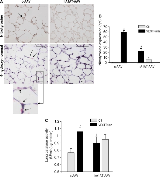
Lung antioxidative effects of hA1AT augmentation in the VEGFR blockade and caspase-3 instillation models. (A–B) Intramuscular transduction of hA1AT-AAV in the SU5416-treated mouse. (A) Representative immunohistochemistry (IHC) of nitrotyrosine (in brown, arrow, upper panel; bar = 50 μm) and 4-hydroxy-nonenal (in red, arrows, lower panel and inset) expression at 3 wk VEGFR blockade. (B) Nitrotyrosine expression (cpf by IHC, mean + SD; *p < 0.05 vs. Ctl #p < 0.05 vs. VEGFR-inh. (C) Intratracheal transduction of hA1AT-AAV in the SU5416-treated mouse. Lung catalase activity 4 wk after VEGFR-inh administration (mean + SEM; *p < 0.05 vs. Ctl #p < 0.05 vs. VEGFR-inh).

