Figure 1.
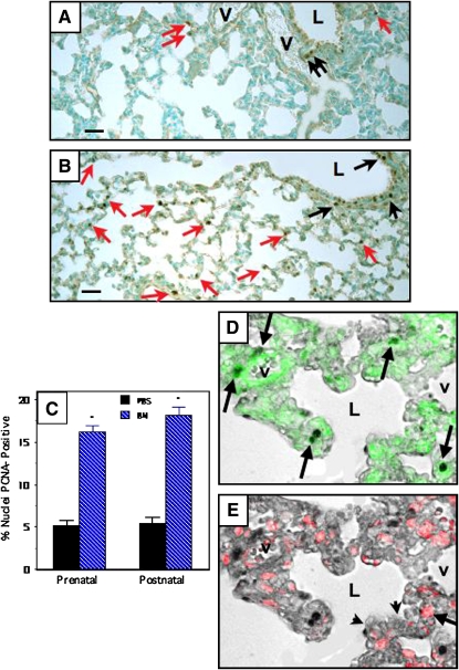
Bombesin (BN) increases alveolar cell proliferation in newborn Swiss-Webster mice as assessed by proliferating cell nuclear antigen (PCNA) immunostaining. Swiss-Webster mice were treated with BN or phosphate-buffered saline (PBS) prenatally (E16–E18) or postnatally (P1–P3), as detailed in Methods. All of the lungs analyzed in this study were from Postnatal Day 14 animals. Immunostaining for PCNA was performed to evaluate the prevalence of alveolar cell proliferation in lung tissue sections. Details of computerized image analysis are given in Methods. (A) Representative section of lung from mouse given PBS postnatally. Note several PCNA-positive nuclei lining airspaces (red arrows). There are also scattered PCNA-positive cells in the airway epithelium (black arrows). (B) Lung from mouse given BN (200 μg/kg) postnatally. There are numerous PCNA-positive nuclei in the alveolar walls (many are indicated by red arrows). There are also multiple PCNA-positive cells in the airway epithelium (some indicated by black arrows). (C) Results of morphometric analyses determining the percentage of nuclei in the alveolar wall that are PCNA-positive. Mice treated with BN either prenatally or postnatally have over a threefold increase in the percentage of PCNA-positive cells (*p < 0.0001 compared with corresponding PBS-treated control group). (D) Immunohistochemistry using lung slides from mice treated with BN demonstrated PCNA labeling by bright-field microscopy merged with SMA immunofluorescence (arrows indicate cells with green cytoplasm and PCNA-positive nuclei). (E) In contrast, PCNA positivity only infrequently colocalized with surfactant protein C (SPC), a marker of alveolar type II cells. Note red cytoplasm indicating SPC immunoreactivity in a cell with PCNA positivity (arrow). Scale bar in lower left corner of (A) and (B) = 50 μm. L = airway lumen; V = vessel.

