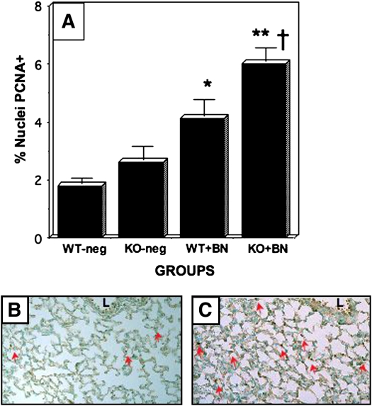
Figure 2.
Bombesin-induced alveolar cell proliferation in GRPR-WT and GRPR-KO mice. GRPR KO and WT mice on a C57BL/6 background were treated with BN or PBS postnatally (P1–P3), and immunostaining for PCNA was performed as described in Figure 1. (A) Results of morphometric analyses determining the percentage of nuclei in the alveolar wall that are PCNA-positive. There was no significant difference in PCNA labeling between untreated WT and untreated KO mice (KO is 1.4-fold greater than WT; p = 0.30). KO mice treated with BN, however, had significantly more PCNA-labeling than BN-treated WT littermates (†p < 0.02). Both WT and KO mice had significant BN-induced responses compared with the corresponding untreated control animals (**p < 0.0001 for KO mouse responses, and *p < 0.005 for WT mouse responses). (B) Representative section of lung from GRPR-WT mouse given BN. Note several PCNA-positive nuclei in developing alveoli (red arrows). (C) Representative section showing PCNA immunostaining of lung from BN-treated GRPR-KO mouse. There are numerous PCNA-positive nuclei in the alveolar walls (many are indicated by red arrows). L = airway lumen.

