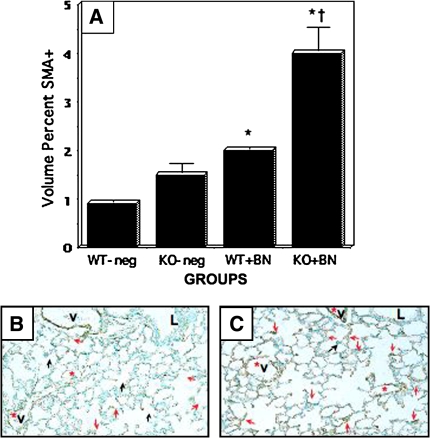
Figure 4.
Bombesin-induced SMA-positivity is enhanced in GRPR-KO mice. GRPR-WT and GRPR-KO mice were treated with BN postnatally (P1–P3), as described in the legend to Figure 2. Immunostaining for SMA was performed as in Figure 3. Black arrows indicate SMA positivity consistent with developing septa. Red arrows indicate SMA-positivity within the interstitium. (A) Results of morphometry assessing the volume percent of SMA-positive cells in developing alveoli. Mice treated with BN have ∼2 to 3-fold increased volume percent of myofibroblasts (*p < 0.0001 compared with the corresponding untreated control group). Bombesin-treated KO mice have significantly more SMA staining than BN-treated WT mice (twofold greater volume percent of SMA; †p < 0.001). (B) Representative section of lung from GRPR-WT mouse given BN. (C) Representative section of lung from GRPR-KO mouse given BN. Compared with Figure 2B, there is an increased prevalence of SMA-positive cells, especially in the alveolar interstitum (red arrows). V = vessel, also indicated by red asterisk.

