Figure 3.
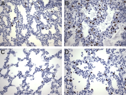
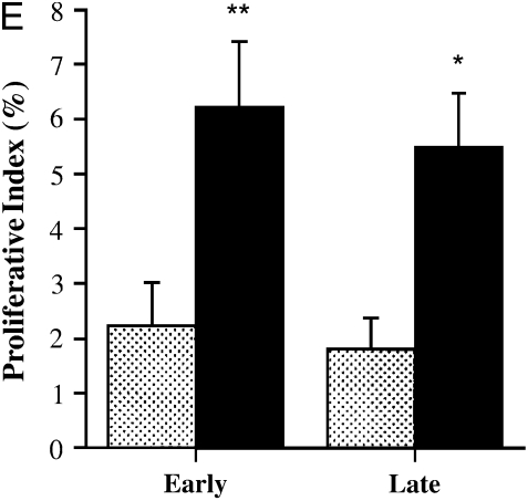
Ki-67 immunolabeling. (A) Early control lung showing scattered Ki-67–positive nuclei within the interstitium and in adlumenal (epithelial) cells (infant born at 24 wk, lived for 2 h). (B) Short-term ventilated lung showing markedly increased numbers of Ki-67–positive nuclei, especially within the interstitium (infant born at 23 wk, lived for 7 d, ventilated). (C) Late control lung showing relatively few Ki-67–positive nuclei in interstitium and epithelium (stillborn at 36 wk). (D) Long-term ventilated lung showing more Ki-67–positive nuclei than late control lungs, evenly distributed over interstitium and epithelium (infant born at 27 wk, lived for 11 wk, ventilated). (E) Lung proliferative index of early control, early (short-term) ventilated, late control, and late (long-term) ventilated infants. Values represent means ± SD of at least five patients per group. Dotted bars, control; black bars, ventilated. *p < 0.05 versus control; **p < 0.01 versus control. Ki-67 immunohistochemistry; DAB with hematoxylin counterstain; original magnification, ×400.


