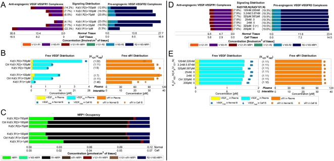
Figure 6. Steady-State Sensitivity to VEGF-Binding Affinities of Cell Surface Receptors: VEGFR1, VEGFR2 and NRP1.
A. Higher VEGF-VEGFR1 affinity and VEGF-VEGFR2 affinity respectively shifted the signaling profile towards anti- and pro-angiogenic complexes. B. Free VEGF levels lowered with increasing VEGF-binding affinity of either VEGFR1 or VEGFR2; while free sVEGFR1 levels rose and fell with increasing VEGF-VEGFR1 and VEGF-VEGFR2 affinity respectively. C. Availability of unbound NRP1 changed inversely with the amount of VEGFR2-VEGF165-NRP1 formed. Free VEGF and sVEGFR1 levels (D), as well as signaling profiles (E), were largely insensitive to NRP1's direct binding-affinity with VEGF. Bracketed percentages in A and D are VEGF-bound fractional occupancies of total VEGFR, averaged (range <0.3%) between normal and calf compartments. ‘+’/‘Ctrl’ = control; ‘Kd(V,R)’ = dissociation constant between VEGF and VEGFR; ‘Kd(V121/V165,N)’ = dissociation constants between VEGF121/VEGF165 and NRP1; ‘R1’ = VEGFR1; ‘sR1’ = sVEGFR1; ‘R2’ = VEGFR2.

