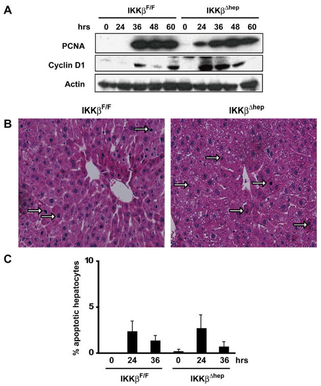
Fig. 1.
IkkβΔhep liver regeneration is accelerated after 70% PH. (A) Precocious PCNA and cyclin D1 expression. Liver lysates were gel separated and immunoblotted. (B) Elevated hepatocyte M.I. Hemotoxylin-and-eosin stained liver sections 48 h post-PH (200×): mitotic hepatocytes (→). (C) Apoptotic IkkβF/F and IkkβΔhep hepatocytes quantified by TUNEL assays (N = 4).

