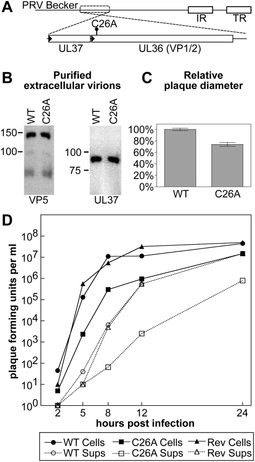
Figure 1. Propagation of PRV with a mutated amino terminus in cultured cells.
(A) Illustration of the PRV-Becker genome with region encoding the UL36 gene (which encodes the pUL36 protein) and neighboring UL37 gene expanded. The position of the codon change resulting in the C26A point mutation is indicated. Promoters are represented as black triangles. IR, internal repeat. TR, terminal repeat. (B) Purified extracellular virions encoding mRFP1-capsids and either a wild-type UL36 allele (WT; PRV-GS847) or UL36 allele encoding the amino-terminal point mutation (C26A; PRV-GS1652) were examined by Western blot analysis for the incorporation of the UL37 protein. The major capsid protein, VP5, was used as a loading control. (C) Comparison of plaque diameters resulting from infection of Vero cells with the virus encoding wild-type UL36 (WT) or mutated UL36 (C26A) allele. Each virus also encodes the mRFP1-VP26 fusion (red capsids). Error bars are standard error of the means (SEM). (D) Propagation kinetics of viruses encoding wild-type UL36 (WT), mutated UL36 (C26A), or a wild-type revertant of the C26A allele (Rev). All viruses also encode the mRFP1-VP26 fusion (red capsids). Infectious virions were detected as plaque-forming units harvested from either the cells (cells) or tissue culture supernatant (sups).

