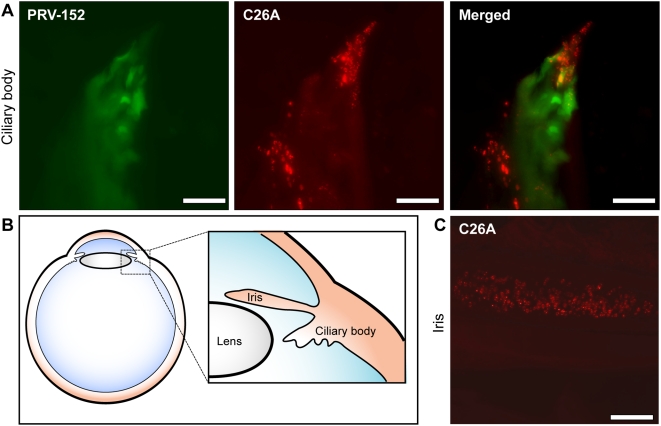
Figure 5. Viral infection in peripheral tissue of the eye.
(A) Images of the ciliary body following co-infection with PRV-152 (diffuse GFP signal) and the C26A virus (punctate RFP signal) in the anterior chamber. Cells infected with both viruses are evident in the merged image. (B) Illustration of the peripheral tissues in the eye (iris and ciliary body) exposed to viral inoculum and imaged in these studies. (C) C26A virus fluorescence from nuclei of cells in the iris following anterior chamber injection. Scale bars = 10 µm.

