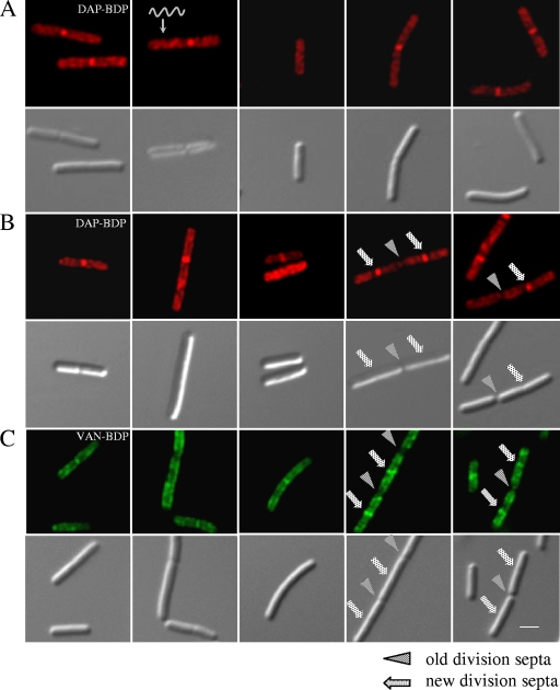
FIG. 2.
Daptomycin-BDP inserts preferentially at new division septa and in a spiral pattern. Fluorescent and DIC micrographs of B. subtilis stained with daptomycin-BDP (DAP-BDP) and vancomycin-BDP (VAN-BDP). (A) B. subtilis W168 treated with daptomycin-BDP at two times the MIC for 10 min (during exponential growth phase). (B) W168 treated with daptomycin-BDP at 10 times the MIC for 10 min. (C) W168 treated with equal amounts of vancomycin and vancomycin-BDP for 20 min. Panels A and B show a spiral localization of daptomycin-BDP and the preferential insertion at newly formed division septa, similar to that of vancomycin-BDP (C). The scale bar represents 2 μm.

