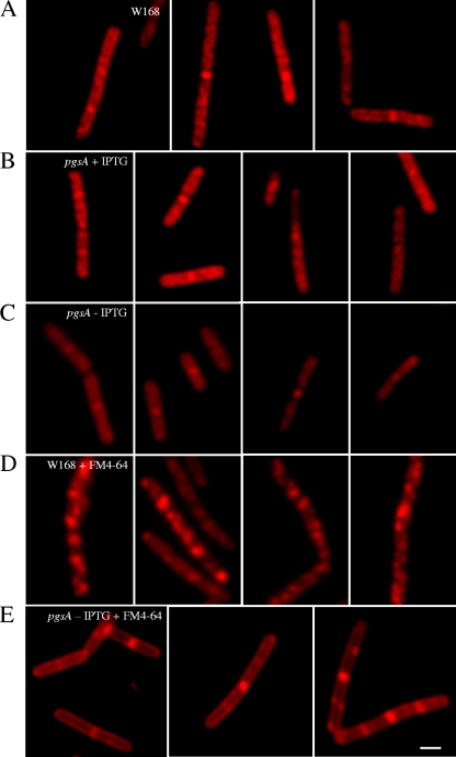
FIG. 4.
Correlation between daptomycin-BDP staining and anionic phospholipid content and distribution. Fluorescent and DIC micrographs of wild-type W168 (A) and pgsA::pMutin grown in the presence (B) or absence (C) of 1 mM IPTG, stained with daptomycin-BDP at two times the MIC for 10 min. Daptomycin-BDP delocalizes when pgsA is not expressed from the IPTG-inducible promoter, suggesting preferential insertion of daptomycin in membrane lipid domains rich in anionic phosphatidylglycerol. W168 and pgsA::pMutin stained with the membrane lipid dye FM 4-64 in the absence of IPTG are shown in panels D and E, respectively. The scale bar represents 2 μm.

