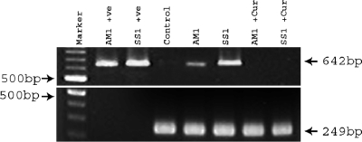
FIG. 4.
Effect of curcumin on H. pylori viability in H. pylori-infected mice. Lanes 1 (second from the left) and 2 represent the amplification of the vacA middle region using DNA from the mouse-colonizing strains AM1 and SS1 with primers VAG-F and VAG-R (Table 1). Lane 3 represents the amplification of bacterium-specific vacA and the mouse-specific GAPDH gene using DNA isolated from the gastric tissue of control mice. Lanes 4 and 5 represent the amplification of vacA and GAPDH using DNA isolated from the gastric tissue of H. pylori-infected mice after 3 weeks of infection. Lanes 6 and 7 indicate the amplification of vacA and GAPDH using DNA isolated from the gastric tissue of curcumin-treated H. pylori-infected mice. +ve, positive control.

