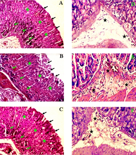
FIG. 5.
Histology of control, H. pylori-infected, and curcumin-treated mouse gastric tissues. Histological sections were stained with hematoxylin and eosin, and photographs were taken at ×10 and ×20 magnifications, respectively. (A to C) Histological appearance of gastric tissues from (A) control mice, (B) mice infected with SS1 for 3 weeks, and (C) mice infected with SS1 and treated with curcumin at 10× magnification. (D to F) Higher-magnification (20×) views of (D) control, (E) SS1-infected, and (F) SS1-infected, curcumin-treated mouse gastric tissues. The gastric mucosal epithelium (black arrows), gastric pits (green stars), gastric glands (green arrows), and inflammatory cell infiltration (black stars) are shown.

