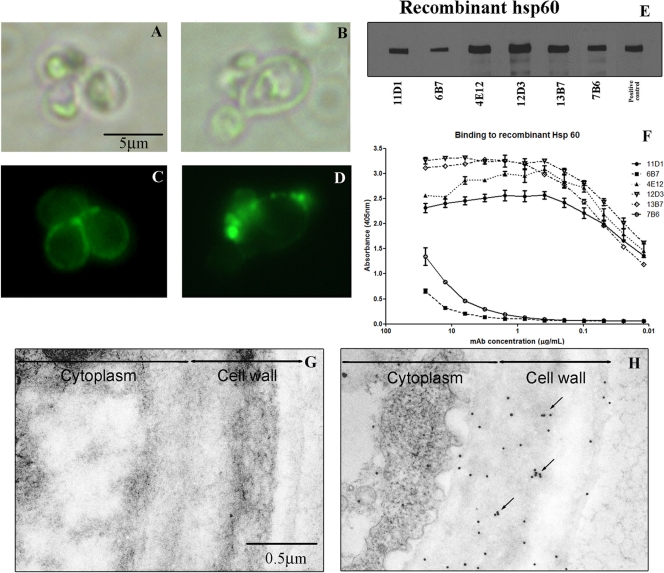
FIG. 2.
MAb-labeled Hsp60 on the cell surface of H. capsulatum var. capsulatum: immunofluorescence and bright-field microscopy showing labeling of H. capsulatum var. capsulatum by MAbs to Hsp60. (A and C) Representative images of diffuse binding with MAb 11D1. (B and D) Punctate binding with MAb 12D3. (E) Immunoblot showing binding of MAbs to rHsp60 protein at 60 kDa. rHsp60 hyperimmune mouse serum was used as a positive control. (F) Representative curves for MAb binding to rHsp60 of yeast cells as determined by indirect ELISA. (G and H) Representative immunogold-labeled transmission electron micrographs for control MAb (G) and MAb 12D3 (H), showing the distribution of Hsp60 on the cell wall and surface of the yeast. Clusters of gold balls labeling Hsp60 consistent with vesicular transport are indicated by arrows.

