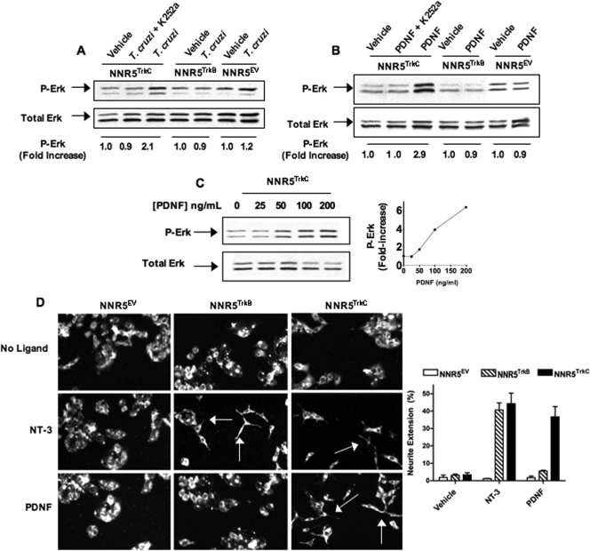
FIG. 5.
T.cruzi/PDNF activates TrkC signaling. (A) Cells were cultured in serum-free medium overnight, treated with the Trk-specific inhibitor K252a (1 μM) for 1 h where indicated, and then treated with 106 parasites or not treated for 12 min at 37°C. Cell lysates were analyzed by Western blotting to evaluate P-Erk and total Erk. (B) Similar to A, but plated cells were treated with PDNF (200 ng/ml) for 12 min. (C) PDNF dose-response treatment of NNR5TrkC as described above (B) with accompanying graph. Phosphorylation (increase) was calculated by scanning densitometry. Each P-Erk band was standardized against its total Erk band and then standardized against vehicle (DMEM), which was arbitrarily set to 1. Experiments in A to C were done twice, with similar results obtained. (D) NNR5TrkC, NNR5TrkB, and NNR5EV cells were plated and cultured for 2 to 3 days with PDNF (250 ng/ml) or NT-3 (100 ng/ml). Cultures were then fixed, probed with anti-neurofilament primary antibody and Alexa 488 secondary antibody, and imaged (magnification, ×20) to visualize neurite extension; arrows point toward neurites. (Inset) Neurite extension was quantified for three individual experiments; error bars represent the standard errors of the means between the three experiments, and 50 or more cells were counted for each condition in each experiment. Neurite extension (percent) is the number of cells with at least one neurite with a length greater than 100% of the diameter of the cell body divided by the total number of cells.

