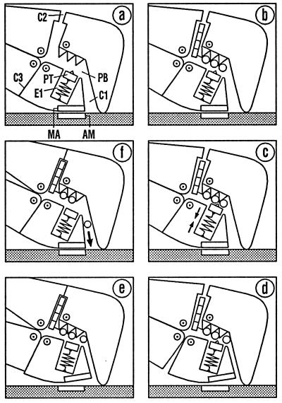
Figure 3.
Events in the vicinity of the phosphate-binding pocket. The diagram is to be read in clockwise sequence. (a–c) Entry of ATP via C2 initiates stretching of the elastic element E1. In d, E1 has succeeded in opening the myosin–actin bond MA/AM. Detachment of the myosin head triggers ATP hydrolysis and closes clefts C2 und C3 (e, without actin). Reattachment starts with the conformation of the free head (e, with actin). Returning to the strong binding state (f) opens cleft C1 so that Pi can leave the head. A further change of the conformation is required for ADP release (see Fig. 4).

