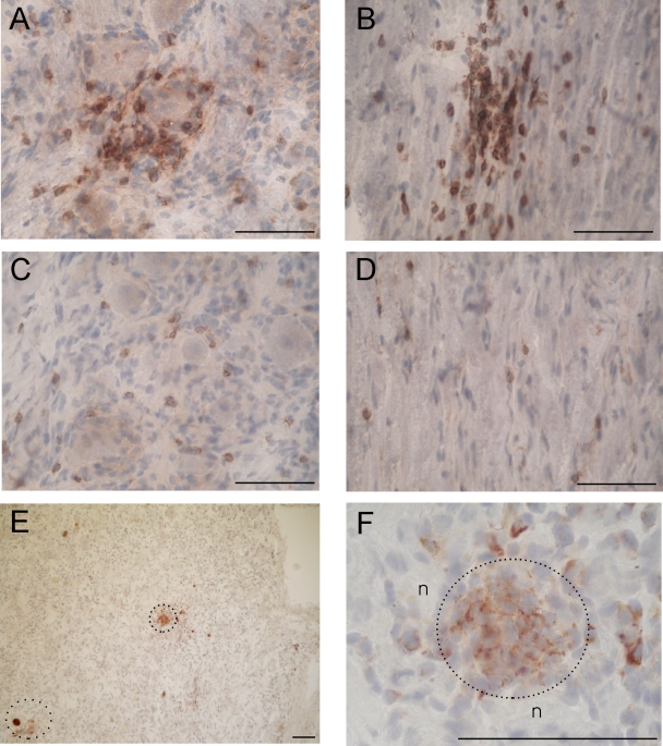
FIG. 5.
Distribution of cytotoxic T cells in the ophthalmic and maxillary divisions and nerves of human TG. CD8+ T cells are arranged around a sensory neuron (A) and clustered in nerve fibers (B) of the maxillary division (V2). Single CD8+ T cells are scattered among sensory neurons (C) and nerve fibers (D) of the ophthalmic division (V1). Also shown are groups of granzyme B-positive T cells in V2 (E; two dotted circles). Panel F shows a higher magnification of the centered cluster (dotted circle; n, sensory neurons [×1,000]). The scale bars are 10 μm in all micrographs.

