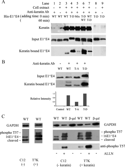
FIG. 6.
16E1^E4 T57D binds keratin more strongly than the 16E1^E4 WT and is protected from proteasomal degradation. (A) Keratins immunoprecipitated from Empigen-soluble fractions of SiHa cells were incubated with 0.4 μg of the His-16E1^E4 WT, His-16E1^E4 T57D (T-D), or a mixture of the two forms. Control immunoprecipitations were carried out in the absence of either cell extract (lane 8) or antikeratin antibody (lane 9). For lanes 6 and 7, T57D and WT His-16E1^E4, respectively, were preincubated with keratin (time point, 0 min) and then the WT (lane 6) and T57D (lane 7) were added 1 h later (time point, 60 min). All samples were analyzed by Western blotting using anti-16E1^E4 antibody and antikeratin antibody (Ab). The T57D form of 16E1^E4 showed greatly enhanced keratin binding compared to that of the unphosphorylated WT 16E1^E4 protein. +, present; −, absent. (B) Empigen-soluble extracts from WT, T57A (T-A), or T57D E1^E4-expressing SiHa cells were immunoprecipitated with a mouse monoclonal antikeratin antibody (L2A1) and analyzed by Western blotting with rabbit polyclonal antibodies against 16E1^E4. T57D was more effectively immunoprecipitated than T57A 16E1^E4 and was enriched compared to the input. The relative intensities of keratin-bound 16E1^E4 WT, T57A, and T57D proteins versus those of the respective inputs are shown in the histogram. (C) An isogenic cell line pair either expressing (T7K) or not expressing (Cl2) keratins (8/18) were infected with rAd16E1^E4 for 24 h. E1^E4 expression was examined by Western blotting, with GAPDH levels being used as a loading control (left panel). The T57-phosphorylated form of 16E1^E4 (phospho T57) is observed only in the keratin-containing cells. The right panel shows results for cells infected with either rAdE1^E4 (WT) or rAdβ-Gal (β-gal) in the presence of the proteasome inhibitor ALLN (10 μM; Calbiochem) for 16 h preharvest. The phosphorylated form of 16E1^E4 was restored in the keratin-negative cell line Cl2 (right panel, second lane) and enhanced in the keratin-positive cell line T7K (right panel, fifth lane) following ALLN treatment.

