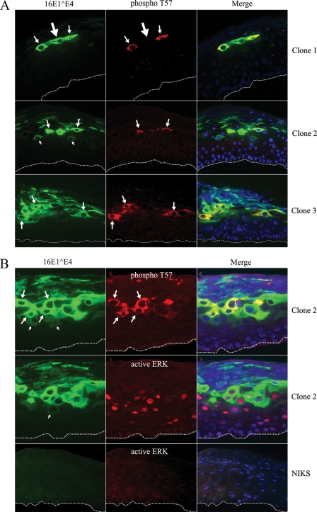
FIG. 7.
T57 of 16E1^E4 is phosphorylated in the intermediate layers in HPV16-containing raft tissue. (A) Organotypic raft cultures from three different HPV16-containing NIKS cell lines (clones 1, 2, and 3) were triple stained for total 16E1^E4 (green), T57-phosphorylated 16E1^E4 (phospho T57; red), and DNA (blue). The cells showing weak cytoplasmic staining of 16E1^E4 are indicated by the small arrows. The cells showing T57 phosphorylation correlating with 16E1^E4 accumulation are indicated by the medium-sized arrows. The cell with abundant 16E1^E4 staining but no detectable T57-phosphorylated E1^E4 is indicated by the large arrow. The dotted lines indicate the position of the basal layer. The images were taken using a 20× objective. (B) Raft cultures from HPV16-containing NIKS cell line clone 2 or HPV16-negative NIKS cells were triple stained with antibodies against total 16E1^E4 (green), T57-phosphorylated 16E1^E4 (red in the upper row), or active ERK (red in the middle and bottom rows) and DNA (blue). The cells showing weak cytoplasmic staining of 16E1^E4 are indicated by the small arrows. The cells showing T57 phosphorylation correlating with 16E1^E4 accumulation are indicated by the medium-sized arrows. The dotted lines indicate the position of the basal layer. The images were taken using a 20× objective.

