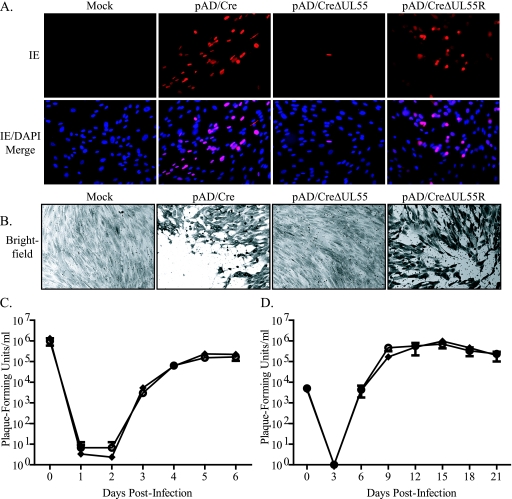
FIG. 2.
Generation and growth kinetics analysis of HCMV from electroporation of BAC plasmids into fibroblasts. NHDF were electroporated in the absence of a BAC plasmid (Mock) or with pAD/Cre, pAD/CreΔUL55, or pAD/CreΔUL55R. At 14 days postelectroporation, IE gene expression was detected via indirect immunofluorescence (A), and 1 week later, the cells were stained with crystal violet to detect plaque formation (B). NHDF were infected at an MOI of 5 (C) or 0.01 (D) with virus generated from electroporation of pAD/Cre (⧫) or pAD/CreΔUL55R (○). Virus-containing supernatant was removed and replaced with fresh medium supplemented with 2% FBS. At time zero and various times postinfection, supernatant was collected and titrated on NHDF. The values represent averages and standard deviations from three separate infections during one experiment.

