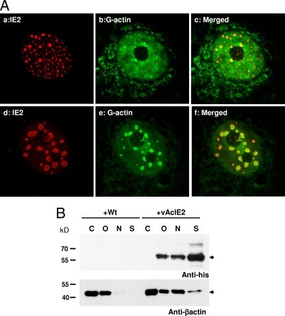
FIG. 4.
IE2 foci interact with G-actin within Vero E6 cell nuclei. (A) Panels a to c show numerous smaller IE2 foci as stained by IF at 16 h p.t. (IE2, red; G-actin, green). Panels d to f show vAcIE2-transduced cells at 20 h p.t. The larger IE2 NBs appeared to contain G-actin (green). (B) Actin redistribution in vAcIE2-transduced cells at 48 h p.t. Cells were fractionated into cytosolic (C), organellar (O), nuclear (N), and cytoskeletal (S) fractions. Western blots detected His-tagged IE2 in noncytosolic fractions and a significant β-actin shift into the nuclear fractions in vAcIE2-transduced cells.

