FIG. 5.
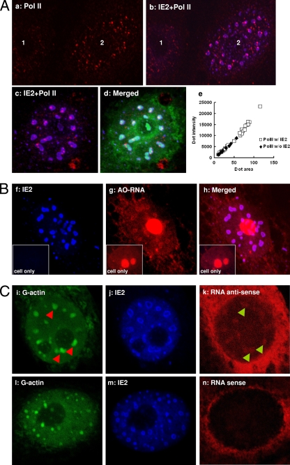
IE2 NBs are active transcription centers. (A) Panels a and b, untransduced (1) or vAcIE2-transduced (2) Vero E6 cells were examined by IF at 16 h p.t. Only nuclei are visible in these photos. Transduced cells showed a greater number of enlarged activated Pol II dots (red), which were associated with IE2 NBs. Panels c and d, an enlarged nucleus showing that activated Pol II, like G-actin (green), locates within the IE2 NBs (blue). IF performed at 20 h p.t. Panel e, a MetaMorph analysis showed that all the larger Pol II dots associate with (w/) IE2, while the Pol II without (w/o) IE2 dots tend to be smaller. (B) vAcIE2-transduced cell nucleus showing that the transcribed RNA, which emits red fluorescence with acridine orange staining (AO-RNA), colocalized with the IE2 NBs. An untransduced cell is shown in each inset as a control. (C) RNA FISH with antisense (i to k) and sense (l to n) probes (red) against the CMV promoter-driven ie2 gene. The antisense probe hybridized with abundant RNA transcripts within IE2 NBs (blue) and colocalized with G-actin (green). The sense probe acted as the negative control. Arrowheads were used to show the relative positions of IE2 NBs and RNA clusters.

