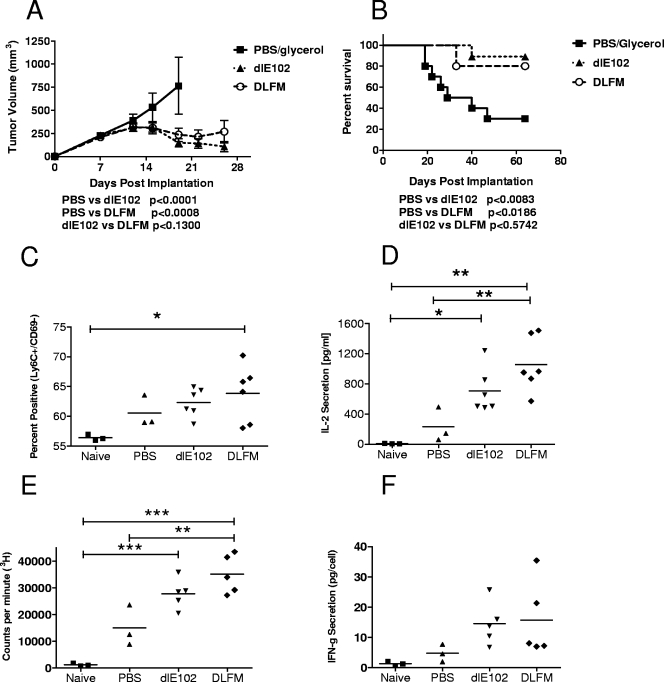
FIG. 6.
Antitumor activity of armed MAV-1 vectors in the highly immunogenic CT26 xenograft tumor model. The DLFM vector was injected into CT26 s.c. tumor-bearing animals, and tumor progression (A) and overall survival (B) were determined. Female BALB/c mice bearing s.c. CT26 tumors (∼150 mm3) were injected twice weekly i.t. with the vehicle (PBS-10% [vol/vol] glycerol), dlE102, or DLFM (1 × 10⁷ PFU in a 50-μl volume). Tumor volume (A) was determined by caliper measurement and expressed as the mean tumor volume (cubic millimeters plus the standard error of the mean, n = 10 per group). Mice whose tumors had been completely eradicated after injection with PBS (n = 3), dlE102, or DLFM (in both groups, n = 6) were rechallenged with CT26 cells. In addition, a naïve control group (n = 3) was also challenged with CT26 cells. Spleens were harvested at 10 days postchallenge, and the percentage of positive CD8+ memory T cells (C) was determined by direct antibody staining and FACS analysis. Spleens were stimulated by coincubation with irradiated CT26 cells, and the levels of IL-2 secretion (D), cell proliferation (E), and antigen-specific IFN-γ secretion (F) were determined. Differences between groups were analyzed by a one-way ANOVA with a Bonferroni correction (*, P < 0.5; **, P < 0.01; ***, P < 0.001); they were not significant for IFN-γ.

