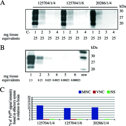
FIG. 1.
(A) Detection of PrPSc by highly sensitive WB analysis in the brain stems and nasal mucosae of three scrapie-positive sheep. Lanes 1 to 4, brain stem, MNC, VNC, and NS, respectively, of the three scrapie-positive sheep; lane C−, MNC of a scrapie-negative sample. The amount of tissue equivalents per lane is indicated in milligrams. All samples were proteinase K digested. (B) WB of a positive brain stem sample (identification number, 125704/1/4/06) serially diluted in negative MNC mucosa: 1 × 10⁻¹ (lane 1), 1 × 10⁻² (lane 2), 1 × 10⁻³ (lane 3), 1 × 10⁻⁴ (lane 4), 1 × 10⁻⁵ (lane 5), and 1 × 10⁻⁶ (lane 6). mw, molecular mass. The amount of tissue equivalents per lane is indicated in milligrams. All samples were proteinase K digested. (C) Graphical representation of the relative amounts of PrPSc signal intensity found in MNC, VNC, and NS in relation to brains of three scrapie-positive sheep.

