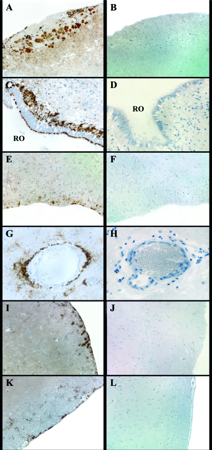
FIG. 6.
Main PrPSc deposition patterns detected in the OS-related brain areas of the scrapie-positive sheep (A, C, E, G, I, and K) and negative control sheep (B, D, F, H, J, and L) examined. (A) Marked fine punctate staining inside the glomeruli of the accessory olfactory bulb (magnification, ×4); (C) recessus olfactorius (RO) of the main olfactory bulb showing a marked subependymal pattern (magnification, ×40); (E) olfactory tract displaying moderate submeningeal and glial PrPSc deposits (magnification, ×10); (G) marked perivascular PrPSc deposition pattern in the frontal cortex at the level of the basal nuclei (magnification, ×40); (I) moderate submeningeal and glial PrPSc deposits in the pyriform lobe (magnification, ×10); (K) apparent subependymal pattern in the hippocampus (magnification, ×10). (B) tissue sections of negative control sheep showing lack of PrPSc immunostaining at the level of the accessory olfactory bulb (magnification, ×4); (D) recessus olfactorius of the main olfactory bulb (magnification, ×40); (F) olfactory tract (magnification, ×10); (H) frontal cortex (magnification, ×40); (J) pyriform lobe (magnification, ×10); (L) hippocampus (magnification, ×10).

