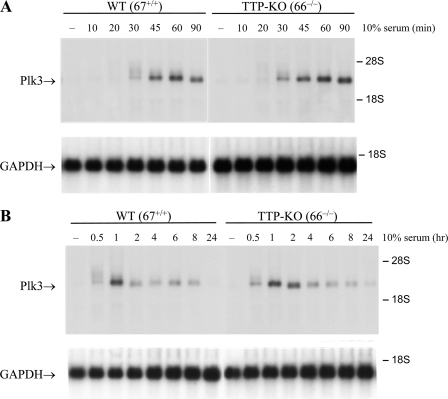
FIG. 1.
Time course of Plk3 mRNA expression after serum stimulation in mouse fibroblasts. Plk3 transcript levels were measured in stable fibroblast cell lines by Northern blot analysis using 10 μg of total cellular RNA and a cDNA probe for Plk3. WT (67+/+) and TTP KO (66−/−) cell lines were serum starved for 16 h in 0.5% FBS and then stimulated with 10% FBS for short (A) and long (B) time courses. The 28S and 18S ribosomal RNAs are indicated with arrows. Equal RNA loading was verified by staining the total RNA with acridine orange (data not shown) and Northern blot analysis of the original blot with a probe for GAPDH (glyceraldehyde-3-phosphate dehydrogenase) (bottom in each case).

