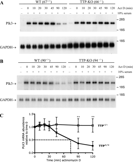
FIG. 2.
Plk3 mRNA stability in TTP-deficient fibroblasts. Plk3 mRNA decay was measured in serum-starved fibroblasts that were stimulated for 60 min with FBS and then treated with actinomycin D for the indicated times. Plk3 mRNA decay was compared in two matched pairs of cell lines: WT 67+/+ and TTP KO 66−/− (A) and WT 90+/+ and TTP KO (94−/−) (B) cells. GAPDH mRNA levels are shown for comparison. (C) The half-life of Plk3 mRNA was examined by averaging data from one of the pairs of fibroblast cell lines (67+/+ and 66−/−); shown are the means of data from four separate experiments. The mean half-life was 74 min for the WT cells in this experiment. Plk3 mRNA levels did not decline sufficiently in the TTP KO cells to calculate the half-life of the Plk3 transcript. The black and white squares represent the WT and TTP KO cell lines, respectively. Error bars represent the standard deviations (SD) derived from four separate experiments, with the Northern blots being quantitated by phosphorimager analysis and normalized for GAPDH mRNA levels. The double asterisks represent P values of <0.05 from a two-tailed Student t test.

