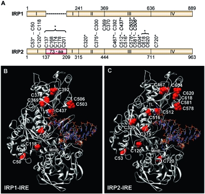
FIG. 1.
Location and structural comparison of cysteine residues in IRP1 and IRP2. (A) Schematic diagram comparing cysteine residues in human IRP1 and IRP2. Conserved cysteines are connected by vertical lines. IRP1 cysteines required for [4Fe-4S] cluster binding are italicized and in boldface (C437, C503, and C506). The 73-aa region is in red, and the asterisk indicates that the five cysteines were mutated in one construct. Asterisks denote mutated cysteine residues. (B and C) Structure for human IRP1 (B) and predicted structure of human IRP2 (without the 73-aa region) (C) bound to the ferritin-H IRE. Both IRPs show IRE contacts at the terminal loop (TL) and midhelix bulge (MHB). Cysteine residues are indicated. The five cysteines located in the 73-aa region of IRP2 (C137, C168, C174, C178, and C201) are not included in the model.

